A new concept for research in the field of stroke involves the participation of neurovascular plasticity after brain injury. At present, there are several cell therapies being investigated that focus on neurorepair. In particular, the administration of endothelial progenitor cells (EPCs) to promote the creation of new blood vessels in the peri-lesional area appears to be a very promising therapeutic strategy to enhance angio-vasculogenic responses during the neuro recovery period.
For a successful treatment it is crucial to ensure the arrival and engraftment of sufficient number of the transplanted cells into the brain. The Neurovascular Research Laboratory at VHIR and the ICMAB-CSIC started a project in 2009 with the working hypothesis that by applying a local magnetic field externally to the brain and by incorporating iron oxide nanoparticles into EPCs, it will be possible to safely guide the arrival of these cells into targeted brain areas where their engraftment will strengthen the endogenous neurorepair mechanisms and thus their angiogenesis potential.
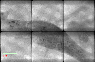
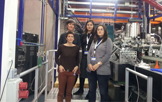
The experiment proposal awarded at the MISTRAL beamline aimed to image whole unsectioned, unstained EPCs containing nanoparticles by perfoming X-ray cryo-tomography. This is one of the three microscopes in the world available to acquire soft X-ray cryo-tomographies.
Following lengthy preparations researchers have obtained the first cryo X-ray tomographies of human endothelial progenitor cells. With this experiment, after imaging reconstruction, they expect to obtain tomographic data sets of 50-60 nm resolution that will allow gathering 3D views of the whole cells allowing a comparative study (size and morphology) of the iron-loaded cells with the pristine cells, as well as between the different EPC subtypes. In addition the researchers aim to determine the spatial bulk distribution of the endosomes/lysosomes containing nanoparticles inside the cells.
The project is currently funded by the Instituto Carlos III-FIS and Ministerio de Economia y Competitividad.
According to the World Health Organization, 15 million persons suffer a stroke worldwide each year. The incidence of this disease in Europe is approximately 200-250 cases every 100,000 inhabitants. It is estimated that at present less than 5% of those patients receive treatment.


