Coccolithophores are widespread marine algae that produce biogenic calcite in the form of minute calcitic scales, known as coccoliths. The coccolith is formed intracellularly, inside a membrane-bound compartment, and upon maturation it is extruded to the cell exterior where many coccoliths are covering the cell surface.
This biomineralization process draws considerable attention since coccolithophores may play an important role in the response of the oceanic ecosystem to predicted global climate change, and since similar changes in the past were recorded in the chemical composition of coccoliths that accumulated in ocean-floor sediments.
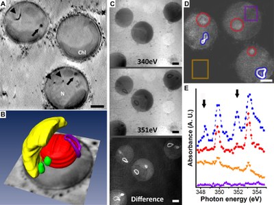
A group of international researchers, led by the Max-Planck Institute of Molecular Plant Physiology (Potsdam, Germany), has used cryo-X-ray tomography at the ALBA Synchrotron for imaging intracellular calcium accumulation in these marine algae. In the first part of this experiment, published in Nature Communications, researchers have discovered high amounts of calcium to be concentrated in membrane-bound compartments that are separate from the coccolith producing compartment. This calcium pool is used for coccolith calcite formation. Inside the compartment calcium is stored as a disordered phase. This finding makes it tempting to speculate that a major fraction of the coccolith calcium is transported as intermediate calcium phase to the site of calcite formation.
"This data provides the first insights on the spatial distribution of calcium in coccolithophorid cells", says André Scheffel, main author of the study. However what is the exact chemical composition of these intermediate phases, and how material is allocated in and out of the compartments is still elusive.
An X-ray microscopy for analysing the sea-bottom
The analyzed cells belong to the model coccolithophore species Emiliania huxleyi that were rapidly frozen and maintained at cryogenic conditions to preserve their intracellular organization.
The cells were imaged at the MISTRAL beamline of the ALBA Synchrotron using X-ray microscopy to acquire two types of data. The first is a tilt-series at the 'water window' energy range. At this X-ray energy the best contrast between carbon-rich intracellular membranes and the water-rich cytoplasm is achieved so the data can be used for 3D reconstruction of cells. The second data set was an energy scan around the Ca L-edge. From these data a complete X-ray Absorption Near-Edge Spectroscopy (XANES) spectrum can be extracted for each pixel in the image, providing information on the concentration of calcium inside intracellular organelles and spectroscopic information on the crystallinity of this Ca-rich phase. In the tomograms of cells all major organelles were visible, as well as intracellular membrane-bound coccoliths in status nascendi. The cells contained distinct intracellular compartments packed with highly absorbing material, which the spectroscopic data showed to be rich in calcium. The XANES spectra collected from multiple Ca-rich compartments were clearly different from the spectra of coccolith calcite and exhibited characteristics of disordered local environment around the calcium atoms.
MISTRAL is one of the seven operational beamlines available at the ALBA Synchrotron. Its X-ray microscopy, together with Bessy (Germany) and ALS (United States), is a powerful tool for analyzing complete cells and for obtaining 3D tomogram, being a ideal complement for other techniques such as the electron microscopy.
Figure: A) Virtual slice of the reconstructed tomogram showing 3 E. huxleyi cells. Immature coccoliths and calcium-rich bodies are marked by arrowheads and arrows, respectively. B) Segmented volume of a tomogram with the nucleus in red, the chloroplast in yellow, the intracellular coccolith in violet and the calcium-rich bodies in green. C) Soft X-ray projections recorded below the Ca L3 edge (top), at the edge (middle) and the absorption difference (bottom). D & E) XANES spectra at the Ca L3,2 edge of the different features shown in D. Scale bars: 1 µm. Picture: Assaf Gal and André Scheffel from the Max-Planck Institute of Molecular Plant Physiology and Andrea Sorrentino from the ALBA Synchrotron.


