ALBA Synchrotron

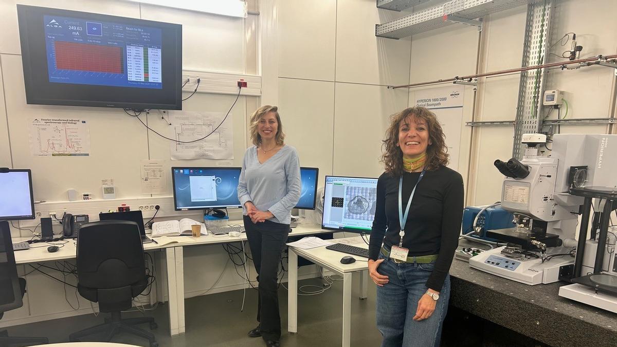
An international team led by Tanja Dučić, scientist at the ALBA Synchrotron and research professor at the University of Belgrade, and Elena González Muñoz, from IBIMA BIONAND Platform and the University of Málaga, has achieved a significant advance in the laboratory study of this highly aggressive brain tumor. The study shows how 3D glioblastoma models respond to a nanocomposite activated and analyzed with synchrotron light.
Glioblastoma is one of the most aggressive brain tumors and carries a very poor prognosis. Its resistance to conventional treatments and its high molecular heterogeneity make it necessary to develop experimental models and analytical tools that allow researchers to study more precisely how tumor cells respond to new approaches based on nanomaterials.
The research investigates patient-derived 3D glioblastoma cell spheroids and compares them with astrocyte spheroid controls, used as a non-tumoral cellular model. The aim was to observe how these complex biological systems respond to a nanocomposite based on carbon dots and riluzole, evaluating the molecular changes that occur across the cellular population after exposure to this formulation.
3D models to better reflect tumor complexity
To study glioblastoma behavior in the laboratory, the team used three-dimensional spheroids: small clusters of cells that grow in a spherical shape. Unlike traditional cell cultures, in which cells develop on a flat surface, these 3D models allow cells to organize themselves in a most similar form to as they do inside a real tumor.
This is important because in a tumor, cells are not isolated: they communicate with one another, are distributed in different layers, and form a complex structure. Spheroids help to better reproduce this tumor architecture and provide a more realistic experimental environment to analyze how glioblastoma responds to potential treatments.
A nanocomposite based on carbon dots and riluzole against the tumor
Carbon dots are nanoscale materials with physicochemical properties that are particularly promising for experimental biomedical applications. This research is part of a line of work developed over more than a decade by Tanja Dučić, scientist at MIRAS beamline of ALBA, focused on the study of experimental glioblastoma models using synchrotron light. Within this framework, the team investigated the effect of a nanocomposite developed by combining carbon dots with the drug riluzole.
The results reveal that this nanocomposite induces clearly detectable molecular modifications in glioblastoma spheroids, particularly in spectral regions linked to nucleic acids, lipids, and proteins. These alterations were significantly more intense in the tumor models than in astrocyte spheroids, where the observed response was considerably more limited.
Synchrotron light to reveal the invisible
The use of synchrotron-based infrared microspectroscopy at the MIRAS beamline of ALBA has been one of the key factors behind the success of this analysis. This scientific infrastructure, where various advanced synchrotron radiation techniques had previously been applied within the framework of the project, has now enabled an approach that acts as a true molecular “supermicroscope". This technology directly and non-invasively detects subtle biochemical changes in proteins, lipids, and nucleic acids, without the need for dyes or chemical markers that could alter the sample.
Thanks to the high intensity and precision of the synchrotron light source, the researchers were able to to map with unprecedented resolution the effects induced by the treatment in the tumor models. The analysis revealed alterations in the structure of cancer cells DNA, signs of oxidative stress associated with damage to cellular membranes, and modifications in the folding of proteins essential for tumor survival, processes that compromise the viability of glioblastoma.
A methodological advance in treatment selectivity in complex systems
One of the most promising findings of the study is the selectivity of the treatment. This approach made it possible to identify clearly differentiated molecular responses between glioblastoma spheroids and astrocytes, providing key insight into the distinct sensitivity of each cell type to exposure to the nanocomposite. While the glioblastoma models showed marked molecular alterations consistent with significant cellular damage, healthy astrocytes exhibited a much weaker response, largely preserving their molecular integrity.
In this context, the study does not propose an immediate clinical application, but rather establishes a high-resolution experimental strategy to analyze how three-dimensional tumor models respond to nanomaterial-based formulations. Overall, the work contributes to the development of advanced methodological tools for evaluating nanocomposites in biological systems that are more complex than conventional cultures, and reinforces the potential of nanotechnology as a pathway toward designing more selective approaches for the study and future treatment of brain cancer.