ALBA Synchrotron

Researchers from the Universidade de Santiago de Compostela, IKERBASQUE, Universidad de Zaragoza-CSIC, Instituto de Ciencia de Materiales de Madrid-CSIC, ESRF, and ALBA Synchrotron (BL29-BOREAS) have led a collaborative research, where they have exploited soft X-ray absorption spectroscopy as a tool for unveiling an electric-field induced crystallographic transformation between the conducting perovskite SrFeO3−δ and insulating brownmillerite SrFeO2.5 at room temperature. The method offers a new approach to design new functional materials with submicrometer spatial resolution.
Reversible structural transformations between perovskite (PV) ABO3−δ and brownmillerite (BM) ABO2.5 (A = Ca2+, Sr2+; B = Fe4+/3+, Co4+/3+) oxides can be induced by topotactic oxygen exchange of reducing/oxidizing conditions. Combining the large oxide-ion conductivity and a small free energy difference between the 4+/3+ oxidation states of 3d transition metal ions enable these topotactic transformations. Herein, the researchers demonstrated that the electric-field produced by a voltage-biased atomic force microscopy (AFM) tip can induce such transformation between the PV SrFeO3−δ and BM SrFeO2.5 at room temperature and with sub-micrometer spatial resolution (Figure 1). Interestingly, after removing the electric field, the structural transformation result in a nonvolatile controlling of the local chemical, electrical, optical, and magnetic properties.
These results, which have just been published in Advanced Functional Materials, open the door for the fabrication of stable ionic-based devices through the electric-field patterning of different crystallographic phases.
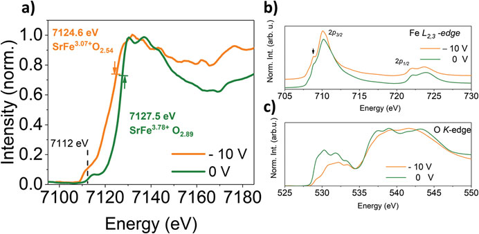
The researchers have demonstrated the topotactic transformation through the changes in Fe oxidation state by exploiting synchrotron X-ray absorption spectroscopy (XAS) at Fe-K, Fe-L2,3, and O K absorption edges, inside and outside an area scanned with a voltage-biased AFM tip. In Figure 2(b) show the isotropic XAS spectra of SrFeOx at Fe-L2,3-edge, for the pristine PV film and for a region scanned at −10 V. The peak at the low energy side of the L3-edge is characteristic of a lower oxidation state of Fe associated to a reduction in the coordination of the transition-metal ion, from octahedral to tetrahedral. The increase of this feature in the region of SrFeO3−δ scanned with the −10 V biased AFM tip supports the occurrence of the PV to BM transformation in this area. The pre-peak in the O K-edge spectra, shown in Figure 2(c), hints at the crystal field splitting that produces a double peak at 530 and 532 eV and its strong suppression after electric field scanning is consistent with an increasing population of tetrahedrally coordinated Fe, as the PV transforms into BM. Overall, the microstructural, Raman, AFM (Topography and conductive probe experiments) and spectroscopic results demonstrate that it is possible to achieve a local stable topotactic PV-to-BM transformation in thin films of SrFeO3−δ, by the direct action of an electric-field-biased AFM tip scanning the bare surface of the film.

Figure: (a) Schematic diagram of the local topotactic transformation between perovskite and browmillerite SrFeO3 achieved by a voltage-biased AFM tip. (b) Optical image of different regions of the film after scanning with −10 V (left). The triangular shadow corresponds to the AFM cantilever. (Right) The scanned areas with the electric field change their color with respect to the conductive PV film background, consistent with a decrease in conductivity.
Acknowledgments:
Financial support from Ministerio de Economía y Competitividad (Spain) under Project Nos. MAT2016-80762-R, MAT2017-82970-C2-R, and PGC2018-101334-A-C22, Xunta de Galicia and the European Union (ERDF) (Centro singular de investigación de Galicia accreditation 2016-2019, ED431G/09), and the Horizon H2020-MSCA-RISE-2016 (No. 734187-SPICOLOST) are acknowledged. The project has received funding from the European’s Union Horizon 2020 research and innovation programme under Grant No. 823717-ESTEEM3. The microscopy work was conducted in the Laboratorio de Microscopías Avanzadas (LMA) at Instituto de Nanociencia de Aragón (INA)-Universidad de Zaragoza. The authors acknowledge the LMA-INA for offering access to their instruments and expertise.
With the collaboration of Fundación Española para la Ciencia y la Tecnología. The ALBA Synchrotron is part of the of the of UCC network from the FECYT and has received support through the FCT-20-15798 project.