ALBA Synchrotron

The structure of cholesterol crystals in atherosclerotic plaques was determined, providing insight on the cellular crystal cholesterol growth mechanisms.
1st September 2018
. Atherosclerosis causes heart attack and stroke and is a major fatal disease in the Western world. The formation of atherosclerotic plaques in the blood vessel walls is the result of low density lipids (LDL) particle uptake, and consequently of cholesterol accumulation in macrophage cells. Excess cholesterol accumulation eventually results in cholesterol crystal deposition, the hallmark of mature atheromas.
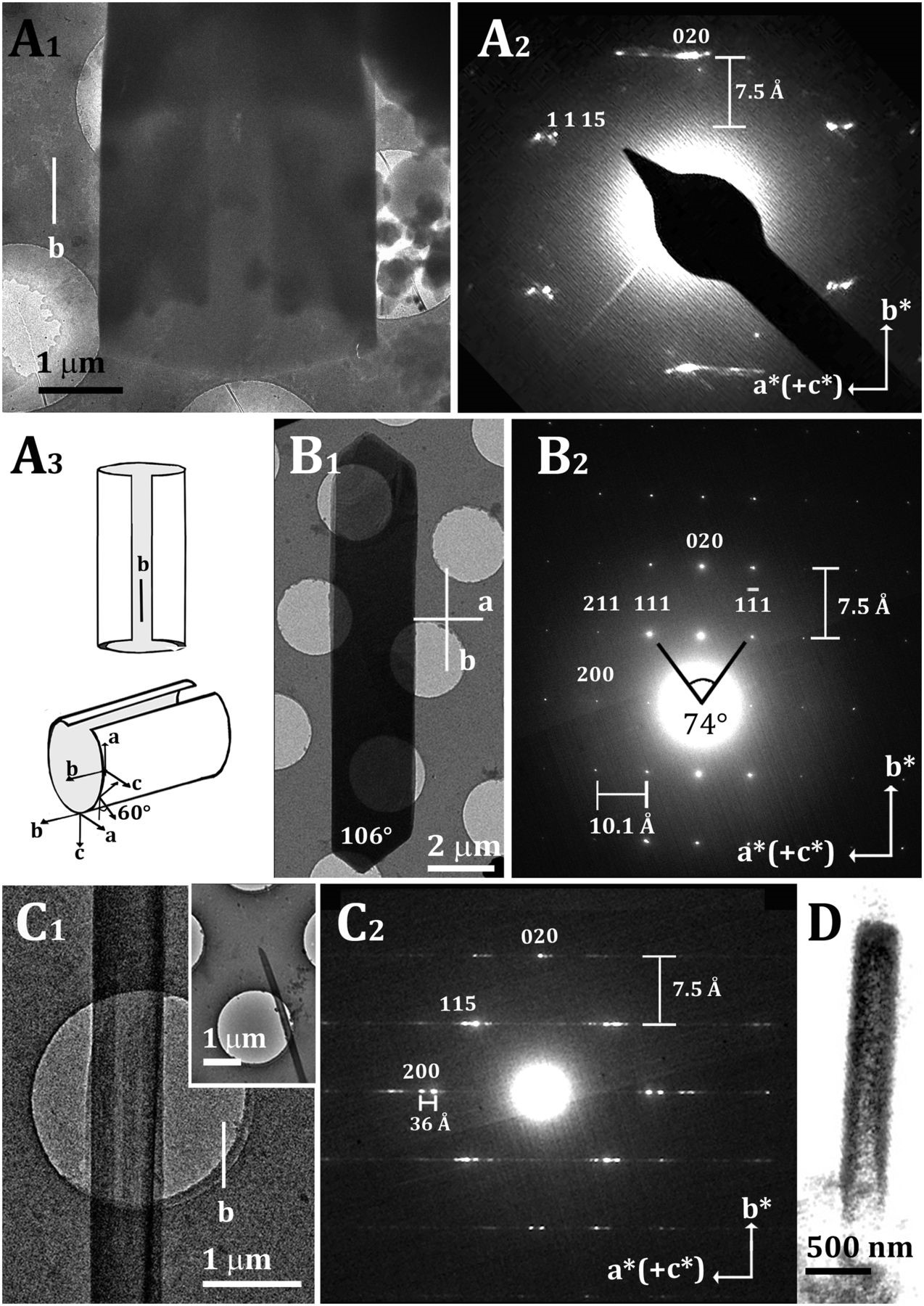
The researchers followed the formation of cholesterol crystals in macrophage cells using correlative cryo-soft X-ray tomography (cryo-SXT) and stochastic optical reconstruction microscopy (STORM) technique. In the initial accumulation stages, small quadrilateral crystal plates associated with the cell plasma membrane are formed. These plates, which may subsequently assemble into large aggregates, match crystals of the commonly observed cholesterol monohydrate triclinic structure. Large rod-like cholesterol crystals form at a later stage in intracellular locations. Using cryotransmission electron microscopy (cryo-TEM) and cryoelectron diffraction (cryo-ED), the researchers show that the structure of the large elongated rods corresponds to that of monoclinic cholesterol monohydrate, a recently determined polymorph of the triclinic crystal structure. These monoclinic crystals form with an unusual hollow cylinder or helical architecture, which is preserved in the mature rod-like crystals. The rod-like morphology is akin to that observed in crystals isolated from atheromas. It is suggested that the crystals in the atherosclerotic plaques preserve in their morphology the memory of the structure in which they were formed. The identification of the polymorph structure, besides explaining the different crystal morphologies, may serve to elucidate mechanisms of cholesterol segregation and precipitation in atherosclerotic plaques.
Cholesterol crystal rods and plates are the evidence of developed atherosclerotic plaques and are associated with plaque rupture and thrombus formation. The structural differences between the two observed polymorphs and their relation to crystal morphology described in this work may now open the path for an investigation, at the molecular level, of cholesterol nucleation and growth. Once the factors that favor the formation of the monoclinic polymorph are understood, mechanistic information on the cellular pathways of cholesterol crystal formation may become evident.

Cryo-TEM images (left) and ED patterns (right) of cholesterol crystals grown in cells and on lipid bilayers. (A) Cryo-TEM image and diffraction of a cholesterol crystal grown in a cell. Proposed open-tube model of the crystal in A1 and A2 is shown in A3. (B) Faceted monoclinic cholesterol monohydrate crystal grown on a supported lipid bilayer. (C) Tubular cholesterol crystal grown on a supported lipid bilayer as in B. (D) Volume rendering of the segmented cryo-SXT data of a crystal from one cell, showing tubular morphology.