ALBA Synchrotron

In a step forward for research into improving outcomes for glioblastoma, researchers from the ALBA Synchrotron, the Institute of Molecular Biology of Barcelona, the Public University of Navarra, and the University of Malaga have come together to successfully synthesize and characterize nanocarriers for the drug riluzole.
Synchrotron light and electron microscopy techniques played a crucial role in providing valuable insights into the mechanism of action of this novel nanoparticle drug delivery system.
Despite an increase in new chemotherapies, the overall prognosis for patients with glioblastoma multiforme (GBM) remains extremely poor, with just 5% of patients surviving for more than five years. This aggressive form of brain cancer is highly resistant to treatment, prompting researchers to explore new treatment avenues. Riluzole, a drug that has already been approved by the FDA to treat amyotrophic lateral sclerosis (ALS), is currently being explored as a treatment for several cancers including GBM. However, there is a need for novel drug delivery methods to enhance riluzole's effectiveness and overcome barriers to targeted therapy, including minimizing harmful side effects in healthy cells, and maintaining the drug's anti-cancer efficacy until it reaches tumor cells.
In this study, which was led by Tanja Dučić, scientist in the MIRAS beamline team at the ALBA Synchrotron, and published in ACS Omega, researchers engineered carbon-based nanoparticles, or carbon dots, made of 2-acrylamido-2-methylpropanesulfonic acid (AMPS). This organic delivery system (AMPS-CDs NPs) showed biocompatibility with glioblastoma cells, and researchers were keen to test its potential to act as a nanocarrier for the drug riluzole.
Several Spanish institutions and researchers collaborated in this project, including Manuel Algarra from INAMAT2 (Institute for Advanced Materials and Mathematics), at the Public University of Navarra; Elena Gonzalez-Munoz, Maria Soledad Pino-González and Juan Soto from the University of Malaga; Pablo Guerra from the Institute of Molecular Biology of Barcelona (IBMB-CSIC); and Tanja Dučić from ALBA.
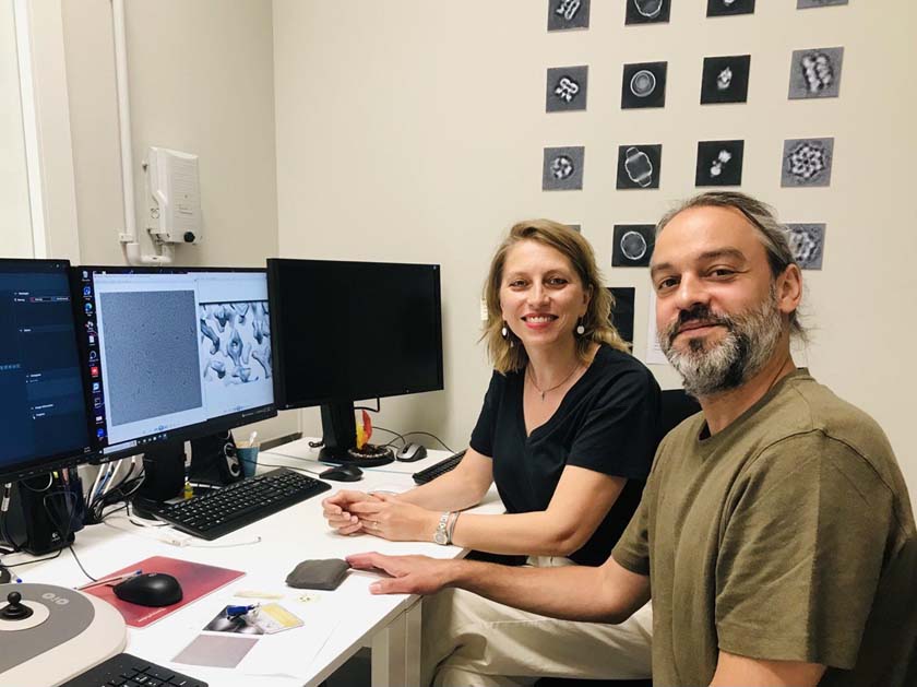
The study demonstrates the successful complementarity between synchrotron light and electron microscopy. By combining the MIRAS beamline and the Cryo-TEM at IBMB-CSIC, part of the Joint Electron Microscopy Center at ALBA (JEMCA), the collaboration achieved its first publication using both instruments. Pablo Guerra, coordinator of the Cryo-TEM, performed the microscope data acquisition. "Using the Cryo-TEM we confirmed the nanoparticles' shape and size, with a diameter of 4.5-5 nm, which was impossible to observe with other methods", says Tanja Dučić.
The nanoparticles were extensively characterized to determine their exact surface composition using techniques that included XPS (X-ray photoelectron spectroscopy) and NMR (nuclear magnetic resonance) spectroscopy, as well as cryo-transmission electron microscopy. The synthesized nanoparticles are covered in sulfonated, carboxylic, and substituted amide groups. These functional groups make the AMPS-CDs potentially suitable nanocarriers for riluzole.
Following the characterization, synchrotron radiation-based Fourier transform infrared microspectroscopy (FTIR) was used to assess the impact of the riluzole-loaded carbon dot nanocarriers on live glioblastoma cells. Researchers were able to observe significant changes in the cancer cells' protein structure, indicating the nanocarrier's ability to effectively deliver riluzole.
The findings show that AMPS-CDs are a promising nanocarrier system for delivering drugs to target proteins within GBM cells, and steps can now be taken to enhance the accuracy and effectiveness of this potential treatment. These measurements benefited from the extremely bright and focused light at ALBA, which allowed the researchers to detect subtle changes in cancer cell composition and biomolecular structure to a level of precision that is crucial when understanding the impact of nanocarriers in binding and releasing the drug.
The promising findings in this study pave the way for further advancements in the treatment of this aggressive brain cancer. Looking to the future, further research could build on this solid foundation to explore the development of targeted treatment options for other types of brain cancer. Further research on non-cancerous cells may provide a more comprehensive understanding of the nanoparticles' overall effects.

An overview of the experimental setup from the carbon dots synthesis, characterisation by EM01-Cryo-TEM and live cells FTIR test. The t-SNE and the PCA analysis show the contribution of individual absorbance of corresponding areas of lipids and proteins.