ALBA Synchrotron

X ray ptychography pushes toward minute scale in situ nanotomography at elevated temperatures, demonstrating the potential of new coherent imaging beamlines for ALBA II.
Researchers from the University of Leeds, Karlsruhe Institute of Technology, Brookhaven National Laboratory, the Paul Scherrer Institute and the ALBA Synchrotron have demonstrated in‑situ ptychographic X‑ray nanotomography methods that capture how materials transform in three dimensions under realistic conditions.

Figure. Tomogram of the formation and recrystallization of a rare metastable polymorph crystal.
(a) Volume renderings showing the formation of a metastable CaCO₃ polymorph crystal (yellow) from amorphous calcium carbonate (ACC, teal) as a function of time and temperature.
(b) Volume renderings showing the recrystallization of the crystal into calcite (red).
Volume renderings are restricted to voxels at one point occupied by the metastable phase.
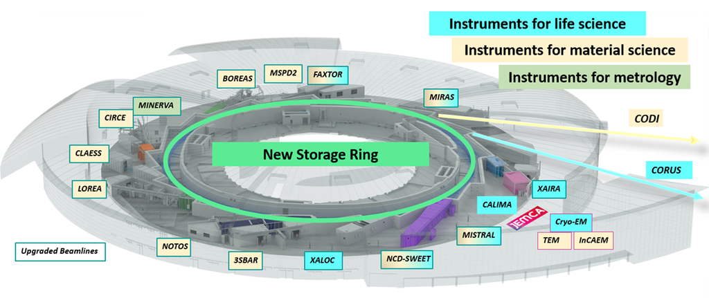
These results showcase the potential of the Coherent Diffraction Imaging (CoDI) beamline, currently under development for the ALBA II upgrade, to deliver operando nanotomography for a broad range of functional materials.
Many technologically important systems, from catalysts to batteries, semiconductors and construction materials, change their internal structure and composition while they operate, but these processes are often too fast, too heterogeneous, or too deeply buried to be followed with conventional imaging methods.
In two recent studies published in Nature Communications, the researchers developed the necessary instrumentation, data acquisition schemes and reconstruction strategies to achieve minute‑scale in‑situ ptychographic nanotomography at temperatures up to 800 °C.
Applied to the of crystallization processes and supported catalyst deactivation, the method reveals heterogeneous pathways, rare transient events and defect evolution that typically remain hidden to bulk or ex‑situ techniques.
CoDI will be a next‑generation coherent imaging beamline at ALBA II, designed to exploit the increased brightness and coherence of the upgraded source for fast, quantitative 3D imaging. By combining ptychographic tomography with advanced sample environments and reconstruction strategies, CoDI aims to enable routine in‑situ and operando studies of complex materials, from energy storage and catalysis to geological and soft‑matter systems.