ALBA Synchrotron

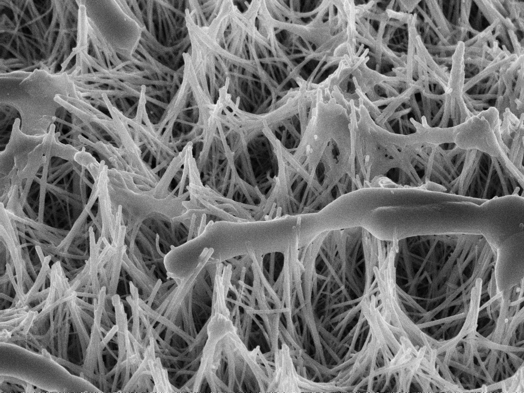
Postoperative infections remain a major challenge in orthopaedic surgery. One promising approach is to engineer the surface of bone-regeneration materials at the nanoscale so they exhibit antibacterial properties. But how exactly do these nanostructured surfaces affect bacteria at the microscopic level? Using advanced imaging techniques at the ALBA Synchrotron, researchers at the Universitat Politècnica de Catalunya (UPC) have now captured how these surfaces interact with bacteria in unprecedented detail.
Bone grafts —a material used to repair, replace, or regenerate damaged bone— have transformed the treatment of many skeletal injuries, but one major challenge remains. Even after a successful operation, bacteria can colonise these materials and cause persistent infections that require prolonged antibiotic treatment or event additional surgery. As antibiotic resistance continues to rise, researchers are exploring a new strategy: what if the bone graft itself could help prevent infection?
One material that attracts growing attention is calcium-deficient hydroxyapatite (CDHA), which resembles the mineral component of human bone and promotes bone regeneration. Under specific processing conditions, CDHA develops a surface made up of densely packed, needle-like crystals only a few nanometres wide.
To a bacterium, this surface is like a bed full of tiny spikes. Previous studies had shown that as the bacterial cell tries to attach, it is forced to stretch across the needle-like crystals resulting in envelope damage or cell death. This make it much harder for bacteria to colonise, but CDHA is more than a hostile physical landscape. Unlike other antibacterial nanostructures made from chemically inert materials such as silicon, gold or titanium, CDHA is a bioactive mineral that continuously exchanges calcium and phosphate ions with its surroundings. How do bacteria respond to a surface that combines a needle-like nanostructure with an active, ion-releasing chemistry?
A new study led by Montserrat Español Pons, Marc Iglesias Fernández and Maria Pau Ginebra Molins - researchers at the Universitat Politècnica de Catalunya (UPC) - has now provided a closer look at this interaction. Working at the MISTRAL beamline of the ALBA Synchrotron, the team combined advanced techniques not only to visualise the bacteria's structure in three dimensions, but also to quantify and map the distribution of calcium inside each bacterial cell.
The researchers exposed colonies of Bacillus subtilis to nanostructured CDHA and then obtained 3D images to examine changes in the bacteria’s overall structure and internal organisation. The images revealed signs of mechanical damage to the bacterial cells: ruptured cell walls, leakage of cytoplasmic contents and the formation of multivesicular bodies—all hallmarks of severe cellular stress. With advanced spectromicroscopy techniques, researchers also found that some bacteria accumulated intracellular calcium after exposure to the CDHA surfaces, consistent with the release of calcium from the material. In other words, the bacteria did not only interact with the nanostructured surface, but also with the calcium released by the material.
Interestingly, the extra calcium did not seem to affect the bacteria’s viability. Bacillus subtilis is naturally tolerant to changes in ion concentrations, which probably explains why it can cope with the additional calcium. These findings suggest that the bactericidal effect observed here is mainly driven by the physical interaction between bacteria and the nanostructured surface, rather than the chemical action of the released calcium.
Nevertheless, visualising this active ion exchange provides valuable insight into how bacteria interact with bioactive bone grafts and opens the door to engineering next-generation materials doped with ions that exert stronger bactericidal activity. By combining alternative ions that induce biochemical damage with optimised CDHA surfaces that maximise mechanical disruption, these findings could guide the design of new biomaterials capable of promoting bone regeneration while further reducing bacterial colonisation.

(a) X-ray image of bacteria after exposure to the nanostructured biomaterial. (b) Three-dimensional reconstruction showing individual bacteria (coloured) alongside crystals of the calcium phosphate material (grey).