ALBA Synchrotron

The CLEAR spectrometer of CLÆSS beamline has reached the operating status and will be open to users for the next call for proposals.
Hard X-ray spectroscopies, such as X-ray emission (XES) or absorption (XAS) spectroscopy, are unique element specific probes of the local electronic and atomic structure. XAS and XES are the two faces of the same phenomena. In XAS, the samples under study are illuminated with photons of selected energies that originate electronic transitions from occupied to empty levels or to the continuum and the evolution of the absorption coefficient as a function of energy gives access to the local electronic and structural properties. In XES, the samples under study are illuminated with photons of energy well above a selected absorption edge, and it is investigated the decay, i.e. the fluorescence signal following the creation of the core hole as a function of the emitted energy to access complementary information on the local electronic, magnetic and structural properties. For example, an atom ionized at the K shell emits several fluorescence lines denoted Kα, Kβ, Lα, Lβ, etc...
In some cases, the energy dependent photon absorption is monitored by measuring the fluorescence emitted by the excited atoms, arising from the refilling of the core hole by an electron on a higher shell. High resolution XAS can be collected by selecting a particular emission line if the emitted photons are discriminated with a sufficiently good energy resolution (in most of the case it is necessary an energy resolution around or below 1eV).
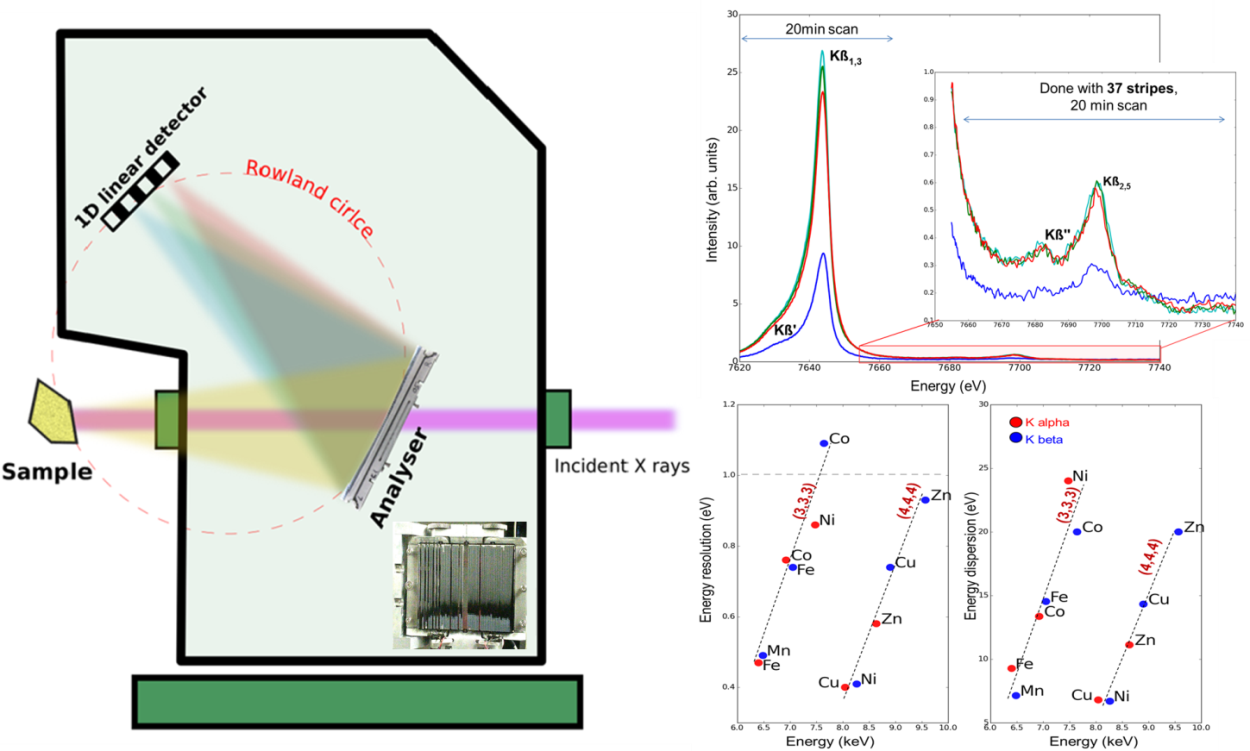
At CLÆSS, the core level absorption and emission beamline of the ALBA Synchrotron, the CLEAR emission spectrometer, conceived by the former ALBA scientist Konstantin Klementiev, has finally reached the operating status. This has been the result of the collaborative effort of scientists, technicians, and engineers under the leadership of Laura Simonelli. The instrument, that has been engineered in-house and manufactured partially-in house and by local companies, allows to energy-analyze the emitted fluorescence and to resolve with good energy resolution (Fig. 1 right, bottom panel) the signals from the different de-excitation channels. Last but not least, thanks to the combined use of a Mythen unidimensional detector and a silicon diced analyzer, it permits the acquisition of the spectrum on a single-shot basis.
In practice this supposes an enormous boost to the scientific possibilities of the beamlines since it will give access to all the complementary information obtainable by investigating the atomic emission lines and, the rather XAS featureless spectra, obtained with the conventional energy integrated mode, become spectra with fine structure containing key information on electronic levels and magnetic properties. Details on the instrument will be published soon.
The CLEAR spectrometer, still under commissioning, is planned to be open to user operation from the next proposals call. We are confident that this instrument will generate a significant advancement in revealing the usually hidden details of the electronic structure of materials.

Figure 1 shows on the left a sketch of the CLEAR spectrometer with, at the lower right, a picture of the presently operating Si(1,1,1) dynamically bent diced analyzer. This instrument is foreseen to work, like the other existing emission spectrometer, in Rowland circle geometry, but, differently from the others, to cover continuously between 2 and 22 keV by means of 4 analyzer crystals (with different crystals reflections) in fully back of forward scattering geometries, exploiting a working wide Bragg angular range (30°-80°). The energy resolution and photon intensity (focal properties) are ensured by the diced analyzer and the analyzer dynamical sagittal bending. While only the Si(1,1,1) analyzer reflection is at the moment available, other analyzers are underway starting from the Si(2,2,0) reflection.

While figure 1 (right) reports an example of recently measured Co Kβ emission lines, Figure 2 shows an example of high resolution absorption spectra collected by selecting the Co Kβ1,3 emission energy and scanning the incoming energy across the Co K-edge. The combination of a unidimensional detector and a diced analyzer permitted the acquisition of the emission spectrum during the incoming energy scan on a single-shot basis (inset in Fig. 2: 2D plot representing the spectral intensity as a function of the emitted (or transferred) and incoming photon energy across the absorption pre-peak). The 2D plots reveal two different final states, not detectable by the classical XAS.