ALBA Synchrotron

X-ray microdiffraction experiments were done in the MSPD beamline of the ALBA Synchrotron to determine the crystalline structure of chladniite.
Cerdanyola del Vallès, 3rd August 2017.
Researchers from the Institute of Materials Science of Barcelona (ICMAB-CSIC), the Autonomous University of Barcelona (UAB), and the National University of Córdoba (Argentina), in collaboration with researchers of the ALBA Synchrotron, have identified a mineral in the region of Córdoba (Argentina), until now only observed in meteorites.
The study, published in European Journal of Mineralogy, affirms that the mineral is chladniite, a complex phosphate belonging to the fillowite group, which contains sodium, calcium, magnesium and iron, and has a trigonal structure. It has been found in a pegmatite, an igneous (magmatic) rock, formed from the slow cooling and solidification of magma.
Chladniite was first encountered in 1993, in the iron meteorite Carlton IIICD. So far, it had only been observed in this type of meteorites, characterized by having undergone fusion and differentiation processes such as igneous rocks. The name of the mineral is in honor of the German physicist and musician Ernst Florens Friedrich Chladni (1756-1827), pioneer in the study of meteorites, who defended its extraterrestrial origin.
The crystalline structure of the mineral has been determined by fixing a very thin section of the rock onto a glass substrate, and by passing synchrotron light through the set, with the technique called through-the-substrate X-ray-microdiffraction. This technique, driven by researchers from ICMAB, began in collaboration with BM25 SPLine, the Spanish beamline of the European Synchrotron Radiation Facility (ESRF) in Grenoble (France), and has subsequently been developed and refined together with researchers from the MSPD beamline of the ALBA Synchrotron.
Two of the innovative features of this characterization technique are both the use of a very fine glass substrate, which allows the easy location of the area to be irradiated, and the use of focalized synchrotron light, which allows enlightening a very small area of the rock and, therefore, the diffraction of individual crystals.

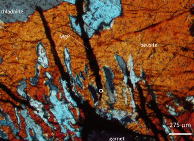
Fig. 1: Cross-polarized-light photomicrograph of the analyzed sample. Greyish blue grains correspond to the complex phosphate chladniite.

Fig. 2: Comparison of 2D frames of the mineral beusite (where chladniite was included) taken by X-ray microdiffraction using the same experimental conditions except for the glass substrate thickness (left: 1.5 mm; right: 0.5 mm).