ALBA Synchrotron

ICMAB researchers lead a study on the toxicity of iron oxide nanoparticles based on the analysis of specific genetic markers linked to nanotoxicity in the C. elegans organisms. Part of the experiment, published in the journal Nanotoxicology, has been carried out at the ALBA Synchrotron, in its infrared light MIRAS beamline. The study suggests that nanoparticles can be captured by intestinal cells in their interior, they can interact with cell lipids, and they can activate cellular mechanisms of oxidative stress.
Cerdanyola del Vallès, 7th August 2017
. Researchers from the Institute of Materials Science of Barcelona (ICMAB-CSIC), led by Anna Roig and Anna Laromaine from the Nanoparticles and Nanocomposites group (N&N), in collaboration with Núria Benseny-Cases, postdoctoral researcher of the , and Stephen Stürzenbaum, from King's College London (UK), have recently published the first article on the MIRAS beamline on the toxicity of nanoparticles.
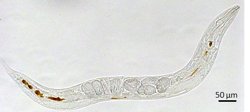
Using the infrared light of the MIRAS beamline, the toxicity of different iron oxide nanoparticles has been studied on C. elegans organisms, the 1-mm-long nematodes used as a model organism for genetic studies, giving that it has its whole genome sequenced. The advantageous characteristics of this worm, such as its transparency, short life cycle (4-5 days), short life (2-3 weeks), ease of operation and low cost of maintenance, among others, are used by the ICMAB group to evaluate the use of nanoparticles for applications in medicine.
The work, in the framework of Laura Gonzalez-Moragas PhD thesis, analyzes changes at the molecular level, influenced by the presence of nanoparticles. The data gathered show that nanoparticles cross the intestinal barrier of C. elegans in processes involving a cell membrane protein, clathrin. In addition, differences in the gene expression are observed depending on the type of nanoparticles to which the worms are exposed. The use of the MIRAS beamline has allowed to determine high levels of oxidation of cell lipids, evidencing tissue oxidative stress after the administration of nanoparticles, which could imply the presence of free radicals or unstable molecules in the organism.
Studies like this demonstrate that by identifying the molecular mechanisms responsible for biological responses observed after exposure to nanoparticles, it is possible to understand and predict their behavior, and optimize their design to achieve low toxicity and safer nanomaterials for humans.
The MIRAS beamline of the ALBA Synchrotron is devoted to infrared spectroscopy and microscopy, covering a range of wavelengths between 1 and 100 μm. It is a very powerful tool to identify the chemical composition of materials at a molecular level using a Fourier transform interferometer. It started commissioning in April 2016 and it opened the doors to academic users through the peer-review panel, operation, in October-2016. The applications of the beamline cover many fields, including materials science, biochemistry, archeology, geology, cell biology, etc.

Fig: Image of the C.elegans showing the nanoparticles inside in brown.