ALBA Synchrotron

An experiment led by Tanja Ducic, scientist at the MIRAS beamline of ALBA, is analyzing live cells with synchrotron-based Fourier Transformed Infrared (FTIR) spectroscopy to identify crucial potential biomarkers in order to target them and to help develop new therapeutic strategies.
Glioblastomas are the most common and deadliest brain cancer in adults, with a life expectancy of only 15 months after diagnosis.
Glioblastomas cells are highly invasive and grow very fast. Despite the scientific community is gaining deeper knowledge about how these cancer cells progress, their mechanisms are not fully understood. Therefore, medical treatment becomes very difficult and challenging.
An international research collaboration, led by scientist Tanja Ducic from the ALBA Synchrotron, is analyzing the different type of live cells extracted from patients and commercial cell line to find out structural and biochemical changes. "The goal is to map different organic compounds in the living cells in order to unveil new therapeutic approaches to target the cancer cells more specifically", says Tanja Ducic.

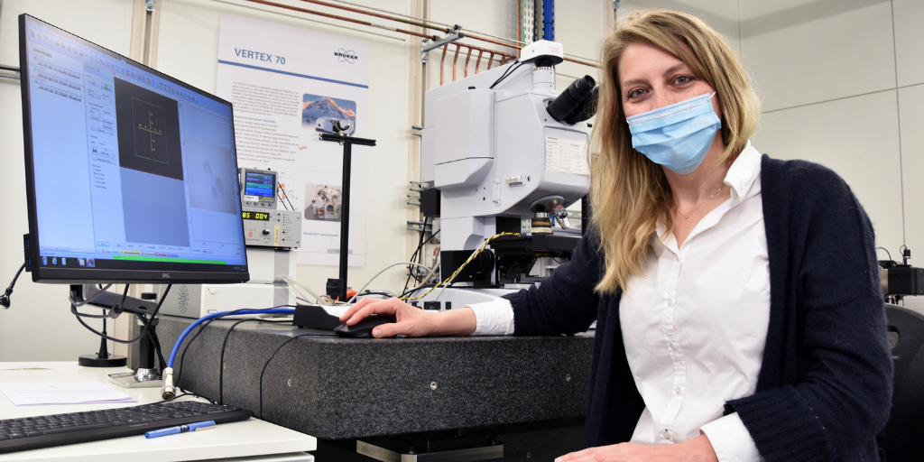
At the left, researcher Tanja Ducic performing the experiment at the MIRAS beamline.
A synergetic and multidisciplinary effort
The team from the Medical Faculty at the University of Göttingen (Germany) were responsible for the sample preparation, collecting primary patient-derived cells and developing stem cells in different stages of the illness.
In order to define a proper set up to study cells in vivo, a new method has been developed by MIRAS team in collaboration with scientists of the Elettra synchrotron (Italy). Living cells are examined in very thin layers of medium, keeping their physiological environment, pH and temperature stable during the measurements. This sample set up preserves cells its natural state, avoiding invasive sample preparation that can cause chemical modifications. The possible effect of riluzole on the biological molecules, as a potential drug on these primary brain cancer cells, is being examined.
Using the , devoted to synchrotron-based Fourier Transformed Infrared (FTIR) spectroscopy, scientists are now identifying differences in the bio-macromolecular compounds of cells. "This technique is very useful to identify the molecular signatures and therefore the chemical composition of biomaterials. So, it brings us the opportunity to see what happens in each single living cell comparing also the possible changes between cell types", states Ducic.
This project is part of a long-term collaboration that already used other synchrotron techniques to shed light on the elemental and structural features of glioblastoma cells established thanks to the financial support of the Union for International Cancer Control (UICC Fellowship no. ICR/2014/339966) and the Northwestern University in Chicago (USA).
"Using the live cells setup we demonstrate our ability to work with patient derived neural cells and information power and possibility which we could obtain by using different complementary synchrotron methods. These new findings may shed new light on the mechanisms of glioblastomas cells behaviour and metastatic potential which will help us to understand how cancer cells changed in comparison to regular cells, giving important indications for future developing more specific treatment strategies", concludes Tanja Ducic.
