ALBA Synchrotron

A new micro-focus experimental station has been developed for the NOTOS beamline in the frame of the InCAEM project. Now available to users, microNOTOS expands the experimental capabilities with microspot X-ray absorption spectroscopy and chemical mapping.
Chemical characterization at the microscale
The primary goal of the new micro-focus experimental station is to perform in situ spectroscopy of samples in miniature reactors, such as those used in transmission electron microscopy (TEM) to study catalytic and electrocatalytic processes. Compatible operando sample environments across complementary characterization tools permit multimodal analysis of complex processes under identical experimental conditions.
In addition, chemical maps of heterogeneous samples can be obtained by scanning the sample position while detecting different X-ray absorption edges, providing valuable information such as composition and oxidation states, with applications in multiple fields such as chemistry, geology, cultural heritage, or environmental studies.
An uncommon geometry, excellent performance
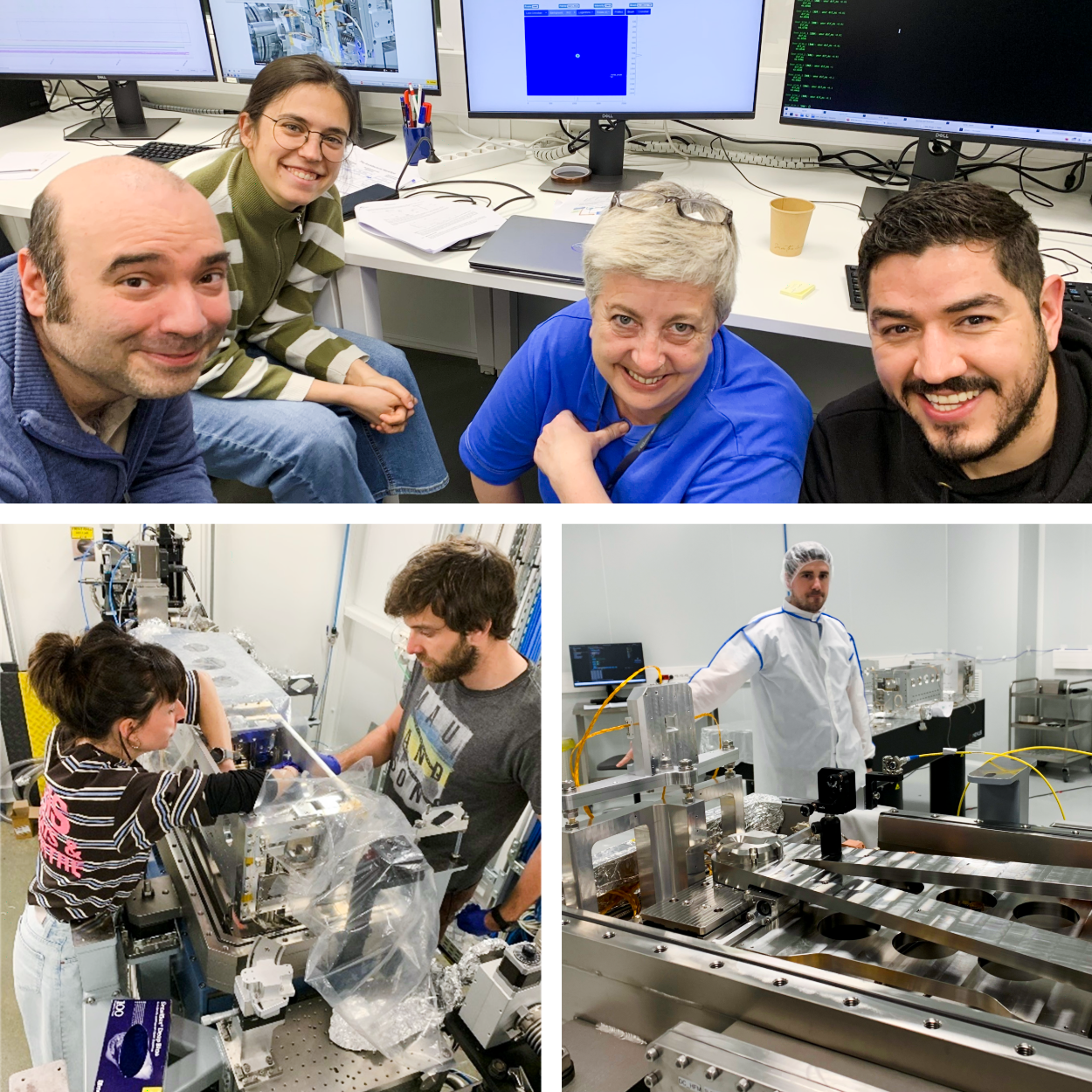
The microNOTOS experimental station has been fully designed, assembled, and tested in-house by a collaboration of scientists, engineers and technicians from different divisions.
The challenge of achieving a micro-focus in an already-built bending magnet beamline was solved designing a pair of hyperbolic mirrors in Kirkpatrick-Baez geometry (KB) mounted on the high precision ALBA nanobender mechanics. The beam can be focused down to a size of 8x7 µm2 (HxV, FWHM), and the photon flux density is increased by 3 orders of magnitude with respect to the unfocused beam.
The experimental station hosts a dedicated sample positioning stage, fluorescence detector, ionization chamber and on-axis sample visualization system. The mirrors can be removed from the beam path in seconds and the full experimental station can be retracted or inserted in the beamline in less than 1 hour.

Overview of the sample positioning stage, fluorescence detector, compact ionization chamber (I₀) and on-axis sample visualization system.

In situ TEM holder mounted on the sample stage.
First measurements with microNOTOS
The performance of microNOTOS was demonstrated measuring CeO2 particles containing 5% wt. CoOx within a fully assembled in situ catalysis TEM holder.
Maps clearly revealed the locations with accumulation of catalyst as well as the position of the metallic resistive microheater, and Co K-edge XANES spectra from microscopic regions could be obtained in a couple of minutes.

Optical micrograph of the central region of a TEM nanoreactor, featuring electron transparent windows (blue), substrate (light green), resistive heater (light orange) and CoOₓ/CeO₂ sample particles (black). The scale bar is 10 µm.

Co K edge spectra from the regions marked by circles on the optical micrograph, with very high and very low sample content in blue and red, respectively. The spectra have been vertically offset for clarity.

X-ray transmission map of the nanoreactor.

Co fluorescence map of the nanoreactor.
InCAEM (In Situ Correlative Facility for Advanced Energy Materials) is part of the Advanced Materials programme by the Spanish Government with funding from the European Union NextGenerationEU (PRTR-C17.I1) and by the Generalitat de Catalunya.